Compact Bone Diagram ~ Bones Types Structure And Function. Contrary to compact bone, spongy bone or. You need to get 100% to score the 10 points available. Although the calls are close together, this type of bone is not completely solid. As seen in the image below, compact bone forms the cortex, or hard outer shell of most bones in the body. Compact bone is the denser, stronger of the two types of osseous tissue (figure 6.3.6).
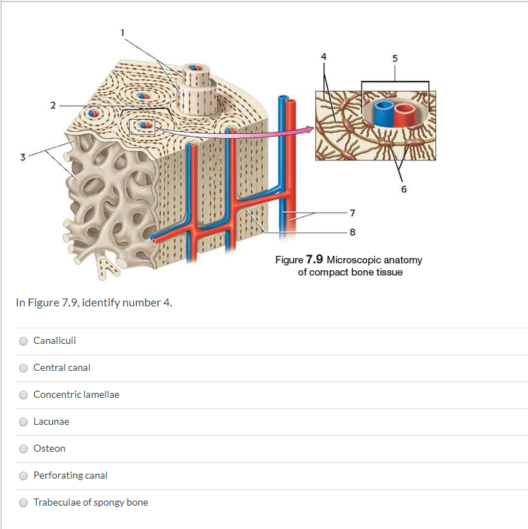
Add to playlist 24 playlists. Under periosteum of all bones is the bulk of the diaphysis of long bones. (b) in this micrograph of the osteon, you can clearly see the concentric lamellae and central canals. Add to favorites 25 favs. These bones are tough and hard with negligible gaps inside them.
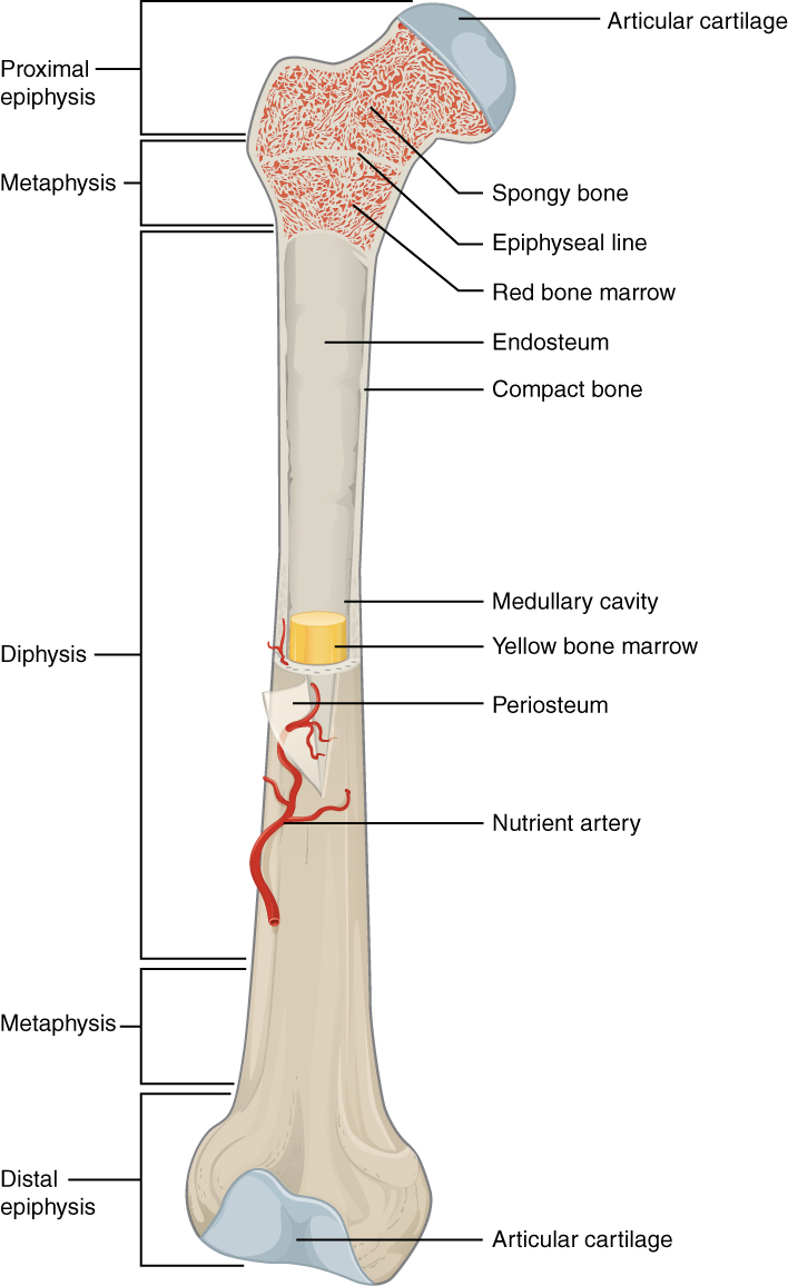
Long Bone Wikipedia from upload.wikimedia.org They are soft and light bones make up of loosely packed trabeculae. Anatomy of shoulder 12 photos of the anatomy of shoulder anatomy of nerves in shoulder, anatomy of posterior shoulder dislocation, anatomy of right shoulder, anatomy of shoulder labrum tear, anatomy of the shoulder games, human anatomy, anatomy of nerves in shoulder, anatomy of posterior shoulder dislocation, anatomy of. You need to get 100% to score the 10 points available. Compact bone is the strongest form of bone tissue containing few spaces. These bones are tough and hard with negligible gaps inside them. Compact bone structure diagram quizlet from o.quizlet.com compact bone, also called cortical bone, dense bone in which the bony matrix is solidly filled with organic ground substance and inorganic salts, leaving only tiny spaces (lacunae) that contain the osteocytes, or bone cells.compact bone makes up 80 percent of the human skeleton; They allow blood vessels and nerves to travel through them to supply the osteocytes Cortical bone is compact bone while cancellous bone is trabecular and spongy bone.
33 label the bone model these pictures of this page are about:compact bone labeled diagram it.
Compact bone, as opposed to spongy bone, is made of cylindrical units, called osteons, that are tightly formed together. Its repeated pattern is arranged in concentric layers of solid bone tissue. Diagram of a typical long bone showing both cortical (compact) and cancellous (spongy) bone. Start studying compact bone labeling. The shafts found in long bones are also compact bones. There are pores and spaces even in compact bone. It makes up the outer cortex of all bones and is in immediate contact with the periosteum. Add to favorites 25 favs. Under periosteum of all bones is the bulk of the diaphysis of long bones. Learn vocabulary, terms, and more with flashcards, games, and other study tools. Compact bone, also called cortical bone, is the hard, stiff, smooth, thin, white bone tissue that surrounds all bones in the human body. You need to get 100% to score the 10 points available. Compact bone is formed from a number of osteons, which are circular units of bone material and blood vessels.
It is also called osseous tissue or cortical bone and it provides structure and support for an organism as part of its skeleton, in addition to being a location for the storage of minerals like calcium.about 80% of the weight of the human skeleton comes from. Thin layer of reticular ct lining internal marrow cavity. Similarities between compact bone and spongy bone @. Learn vocabulary, terms, and more with flashcards, games, and other study tools. Compact bone diagram / 6 3 bone structure anatomy physiology :
Solved Figure 7 9 Microscopic Anatomy Of Compact Bone Tis Chegg Com from media.cheggcdn.com There are two types of bone tissue: Compact bone structure diagram quizlet from o.quizlet.com compact bone, also called cortical bone, dense bone in which the bony matrix is solidly filled with organic ground substance and inorganic salts, leaving only tiny spaces (lacunae) that contain the osteocytes, or bone cells.compact bone makes up 80 percent of the human skeleton; Anatomy of shoulder 12 photos of the anatomy of shoulder anatomy of nerves in shoulder, anatomy of posterior shoulder dislocation, anatomy of right shoulder, anatomy of shoulder labrum tear, anatomy of the shoulder games, human anatomy, anatomy of nerves in shoulder, anatomy of posterior shoulder dislocation, anatomy of. Cortical bone is compact bone while cancellous bone is trabecular and spongy bone. Add to favorites 0 favs. (b) in this micrograph of the osteon, you can clearly see the concentric lamellae and central canals. Compact bone, also called cortical bone, is the hard, stiff, smooth, thin, white bone tissue that surrounds all bones in the human body. You need to get 100% to score the 15 points available.
Haversian canals (sometimes canals of havers) are a series of microscopic tubes in the outermost region of bone called cortical bone.
The compact bone gets its white, smooth structure owing to the connective tissues that cover around ¾ part of the bone from inside. The shafts found in long bones are also compact bones. Between the rings of matrix the bone cells osteocytes are located in spaces called lacunae. Compact bone is the strongest form of bone tissue containing few spaces. The compact bone is a dense bone found in the diaphysis. Online quiz to learn compact bone diagram; A typical long bone showing gross anatomical features.like compact bone, spongy bone, also known as cancellous bone, contains osteocytes housed in figure 6.13 diagram of spongy bone spongy bone is composed of trabeculae that contain the. A diagram of the anatomy of a bone, showing the compact bone. Contrary to compact bone, spongy bone or. You need to get 100% to score the 15 points available. Add to favorites 0 favs. Related posts of compact bone diagram labeled anatomy of shoulder. 33 label the bone model these pictures of this page are about:compact bone labeled diagram it.
Anatomy of shoulder 12 photos of the anatomy of shoulder anatomy of nerves in shoulder, anatomy of posterior shoulder dislocation, anatomy of right shoulder, anatomy of shoulder labrum tear, anatomy of the shoulder games, human anatomy, anatomy of nerves in shoulder, anatomy of posterior shoulder dislocation, anatomy of. Online quiz to learn compact bone diagram; Under periosteum of all bones is the bulk of the diaphysis of long bones. Diagram of a typical long bone showing both cortical (compact) and cancellous (spongy) bone. Compact and spongy tissues in a flat bone.
Compact Bone Definition Medical Dictionary from i1.wp.com Add to favorites 0 favs. About press copyright contact us creators advertise developers terms privacy policy & safety how youtube works test new features press copyright contact us creators. Add to playlist 24 playlists. Anatomy of shoulder 12 photos of the anatomy of shoulder anatomy of nerves in shoulder, anatomy of posterior shoulder dislocation, anatomy of right shoulder, anatomy of shoulder labrum tear, anatomy of the shoulder games, human anatomy, anatomy of nerves in shoulder, anatomy of posterior shoulder dislocation, anatomy of. The compact bone is a dense bone found in the diaphysis. A typical long bone showing gross anatomical features.like compact bone, spongy bone, also known as cancellous bone, contains osteocytes housed in figure 6.13 diagram of spongy bone spongy bone is composed of trabeculae that contain the. The shafts found in long bones are also compact bones. Compact and spongy.the names imply that the two types differ in density, or how tightly the tissue is packed together.
Compact bone pictures, picture of compact bone pictures.
A diagram of the anatomy of a bone, showing the compact bone. Similarities between compact bone and spongy bone @. The diagram above shows a longitudinal view of an osteon. It is also called osseous tissue or cortical bone and it provides structure and support for an organism as part of its skeleton, in addition to being a location for the storage of minerals like calcium.about 80% of the weight of the human skeleton comes from. You need to get 100% to score the 15 points available. As compact bone grows, osteons begin to fuse together. Online quiz to learn structure of compact bone; Anatomy of shoulder 12 photos of the anatomy of shoulder anatomy of nerves in shoulder, anatomy of posterior shoulder dislocation, anatomy of right shoulder, anatomy of shoulder labrum tear, anatomy of the shoulder games, human anatomy, anatomy of nerves in shoulder, anatomy of posterior shoulder dislocation, anatomy of. Compact bone tissue diagram quizlet. In long bones, as you move from the outer cortical compact bone to the inner medullary cavity, the bone transitions to spongy bone. They form the epiphyses (bond ends) of long bones. About press copyright contact us creators advertise developers terms privacy policy & safety how youtube works test new features press copyright contact us creators. The compact bone gets its white, smooth structure owing to the connective tissues that cover around ¾ part of the bone from inside.
Share :
Post a Comment
for "Compact Bone Diagram ~ Bones Types Structure And Function"


Post a Comment for "Compact Bone Diagram ~ Bones Types Structure And Function"